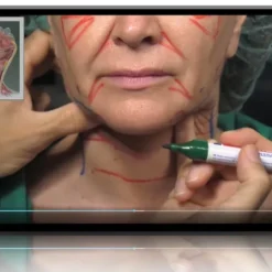
Cutaneous Basic course facial ultrasound 2024
Original price was: €180.00.€51.99Current price is: €51.99.
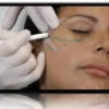
The facial ultrasound basic course by Peter Velthuis and Leonie Schelke introduces imaging physics, probe control and Doppler, then scans every major facial region to prepare injectors for safer, image-informed treatment.
- Ultrasound physics, transducer handling & machine settings
- Temple, supraorbital, nose, midface, lips, chin & jawline scans
- Tissue recognition examples & 19 PDF handouts
- Instant access · unlimited access in time

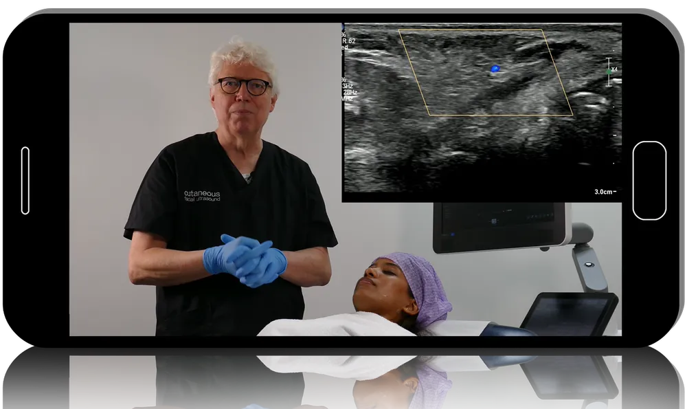
A short look inside — full masterclass unlocked at checkout.
Read facial ultrasound images confidently before guiding a filler needle
The facial ultrasound basic course from Cutaneous teaches transducer handling, depth, gain and focus settings, Doppler use and equipment choice, then scans the temple, supraorbital area, nose, midface, lips, chin and jawline so injectors can recognise skin layers, vessels and filler deposits on screen.
-
27listed lessons
-
19PDF handouts
-
1h 19mlisted teaching
-
2expert instructors
The programme
Physics & technical basics of imaging
After an introduction, the course explains how an ultrasound image is formed and what the main technical controls do. Clear grounding in these principles makes later regional scans far easier to interpret.
Transducer handling & axes of movement
Dedicated lessons show how probe position, pressure and movement along different axes change what appears on the screen. Practising these manoeuvres is the foundation of a reproducible facial examination.
Machine settings, Doppler & equipment choice
Depth, gain and focus are adjusted on screen to demonstrate their effect on image quality. Basic Doppler use for locating vessels and a comparison of available ultrasound devices help clinicians plan their own setup.
Recognising tissue in the facial ultrasound basic course
Example images teach the appearance of different tissues and layers under the probe. This visual vocabulary is what allows a practitioner to separate native anatomy from injected product during a scan.
Regional scans from forehead to jawline
Short region-by-region lessons cover the temporal and supraorbital zones, nose, midface, lips, chin and jawline. Each one shows the structures worth identifying before treatment, closing with an outro by Peter Velthuis.
Who it’s for
- Aesthetic doctors start using ultrasound to add precision to filler procedures
- Dermatologists and radiologists build a base in high-resolution facial scanning
- Plastic surgeons and advanced injectors prepare for ultrasound-guided filler placement and complication work
The consult
Do I need previous ultrasound experience?
No. This is the foundational level, starting with imaging principles, probe control and machine settings before moving into facial regions.
Why are fewer than 27 lesson titles shown?
The package is described as 27 lessons plus 19 PDFs, but the curriculum names only 16 video titles. Expect a compact video set totalling 1 hour 19 minutes supported by the PDF documents.
Does it teach ultrasound-guided injection in depth?
It lays the groundwork for guided injection by teaching examination of each facial region. Hands-on scanning practice under supervision remains necessary before relying on ultrasound clinically.
Who teaches the course?
The lessons are presented by Peter Velthuis and Leonie Schelke of Cutaneous, a group combining aesthetic medicine, dermatology and radiology expertise.
Is access limited to a set period?
No. The videos and PDFs become available the moment payment is completed and remain yours without a time limit.
Do I get CME credit or a certificate?
No. Derma Academy provides the course files only — no CME or CE credits, MOC points or certificates of completion are included.
The spec label
| Format | Self-paced video course — 1 module with PDF handouts |
|---|---|
| Listed duration | 1 hour 19 minutes |
| Faculty | Peter Velthuis · Leonie Schelke |
| Level | Foundational facial ultrasound |
| Techniques | Probe handling · depth, gain & focus · Doppler · regional facial scanning |
| Training models | Narrated live scans · example images · PDF documents |
| Discipline | Aesthetic Medicine · Facial Ultrasound |
| Delivery | Instant access · unlimited access in time |
| Author | Peter Velthuis, Leonie Schelke |
|---|
Related products
Aesthetic Medicine
Aesthetic Medicine
Aesthetic Medicine