2024 Practical Head & Neck Imaging Mimics and Case-based Review

Original price was: €610.00.€69.99Current price is: €69.99.
The head and neck imaging review combines anatomy lectures, “met or mimic” sessions and case reviews from five neuroradiologists to help clinicians interpret CT, MRI and PET-CT across the whole head and neck region.
- Temporal bone, orbit, sinonasal & skull base anatomy and pathology
- Suprahyoid and infrahyoid spaces, nodes, HPV oropharyngeal cancer & perineural spread
- Parathyroid imaging, CSF leaks, PET-CT pitfalls & ENT emergencies
- Instant access · unlimited access in time

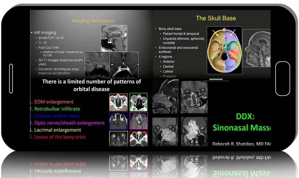
A short look inside — full masterclass unlocked at checkout.
Separate true head and neck pathology from its imaging look-alikes
The head and neck imaging review pairs anatomy lectures on the temporal bone, orbit, skull base, suprahyoid and infrahyoid neck with “met or mimic” sessions and case-based reviews covering sinonasal masses, oropharyngeal cancer with an HPV update, perineural spread, lymphadenopathy, parathyroid localisation, CSF leaks, PET-CT and non-traumatic ENT emergencies.
-
25recorded lectures
-
17h 40mlisted teaching
-
5neuroradiology faculty
-
Unlimitedcourse access
The programme
Temporal bone, inner ear & cerebellopontine angle
Start with CT anatomy of the ossicular chain and facial nerve canal, then move to otomastoiditis, cholesteatoma and their intracranial complications. Later talks review lesions of the internal auditory canal and cerebellopontine angle and close with a temporal bone tumour case review.
Orbit, sinuses & skull base
Orbital anatomy and a case-driven pathology session sit alongside lectures on inflammatory sinus disease, fungal sinusitis and the differential of sinonasal masses. Skull base teaching contrasts metastases with their mimics and outlines how imaging localises the bony defect in CSF leaks.
Spaces of the neck in the head and neck imaging review
Spatial anatomy of the suprahyoid neck — parapharyngeal, pharyngeal mucosal, parotid and carotid spaces — frames how lesions are categorised. Infrahyoid, nasopharyngeal, oral cavity and submandibular sessions extend the same approach lower in the neck.
Head and neck cancer, staging & surveillance
Lectures examine oropharyngeal neoplasms and the rise of HPV-related disease, nasopharyngeal carcinoma spread patterns, cervical nodal assessment and perineural tumour extension along cranial nerves. Separate sessions tackle post-treatment recurrence versus mimic and the strengths and pitfalls of PET-CT.
Glands, nerves & vascular lesions
Parathyroid localisation with ultrasound, CT, nuclear medicine and PET tracers is covered in depth, as is pleomorphic adenoma and its look-alikes. Additional talks map the trigeminal nerve from origin to end organ, classify vascular anomalies and review pituitary tumours against their mimics.
Emergencies & error avoidance
A case-based session on non-traumatic ear, nose, throat and neck emergencies focuses on infections that demand prompt recognition. Across the programme, faculty highlight frequently missed diagnoses and the imaging features that change management.
Who it’s for
- General radiologists build confidence reading head and neck CT, MRI and PET-CT studies
- Neuroradiologists refresh current topics from skull base to oropharyngeal cancer staging
- Otolaryngologists understand how imaging findings shape surgical planning and follow-up
- Radiation oncologists and neurosurgeons sharpen recognition of nodal disease, perineural spread and skull base lesions
The consult
Is this a surgical or an imaging course?
It is purely diagnostic imaging. There are no operative videos; every lecture centres on reading CT, MRI, ultrasound, nuclear medicine or PET-CT studies of the head and neck.
How much of it is case-based?
Four sessions are formal case reviews — orbital pathology, PET-CT in oncology, ENT emergencies and temporal bone tumours — and several “or mimic” lectures also work through examples rather than pure didactics.
Who teaches the lectures?
Five neuroradiologists share the programme, each giving four to six talks. Patricia Rhyner leads the temporal bone and emergency sessions, C. Douglas Phillips covers the orbit and parathyroid, Lawrence Ginsberg focuses on oncology, while Deborah Shatzkes and Richard Wiggins handle sinonasal, vascular, nodal and cranial nerve topics.
Is it relevant to an aesthetic or plastic surgery practice?
Only indirectly. It suits clinicians who order or interpret head and neck imaging; it does not teach cosmetic procedures.
Can I watch at my own pace?
Yes — all 25 lectures are released to you as soon as payment is confirmed and stay available indefinitely.
Do I get CME credit or a certificate?
No. Derma Academy provides the course files only — no CME or CE credits, MOC points or certificates of completion are included.
The spec label
| Format | Recorded lecture series — 25 talks |
|---|---|
| Listed duration | 17 hours 40 minutes |
| Faculty | Rhyner · Phillips · Ginsberg · Shatzkes · Wiggins |
| Level | Intermediate to advanced diagnostic imaging |
| Techniques | CT · MRI · ultrasound · nuclear medicine · PET-CT |
| Training models | Didactic lectures · case-based review sessions |
| Discipline | ENT · Head & Neck Imaging |
| Delivery | Instant access · unlimited access in time |
| Author | Patricia Rhyner, C. Douglas Phillips, Lawrence E. Ginsberg, Deborah R Shatzkes, Richard H. Wiggins |
|---|
Related products

Aesthetic Medicine
From Lab to Practice: A Surgical Guide to Hair Transplantation