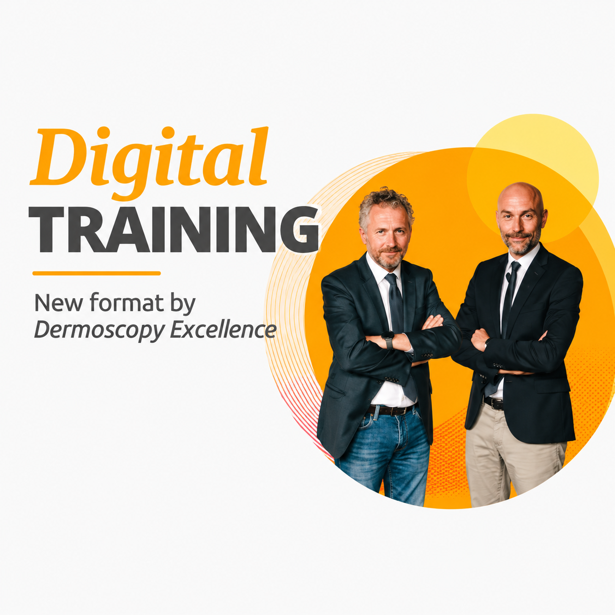
Digital TRAINING from Dermoscopy Excellence


€120.00
The Argenziano Lallas dermoscopy course offers ten hour-long studio modules covering tumours, special sites, monitoring and general dermatology, with more than 500 clinical cases discussed by the two tutors.
- 10 modules · 20 topics · about 10 hours
- Naevi, melanoma, BCC, SCC, spitzoid & amelanotic lesion rules
- Face, scalp, acral & nail lesions, digital monitoring, trichoscopy
- Instant access · unlimited access in time

Learn dermoscopic criteria from two leading teachers, one hour at a time
The Argenziano Lallas dermoscopy course alternates two International Dermoscopy Society presidents across ten studio-filmed modules, moving from light modes and structure–histology correlation through naevi, melanoma, keratinocyte cancers and benign tumours to special sites, spitzoid and amelanotic rules, digital monitoring, inflammatory and infectious disease and trichoscopy.
-
10one-hour modules
-
20topics
-
500+clinical cases
-
2expert tutors
The programme
Instruments, light & the dermoscopic vocabulary
The opening module explains the history of the technique, device choice and polarised versus non-polarised light. It then builds the basic structure vocabulary and links each pattern to what the pathologist would see under the microscope.
Naevi, melanoma & keratinocyte cancers
Fundamental naevus and melanoma patterns come first, followed by basal cell and squamous cell carcinoma. Seborrhoeic keratoses, solar lentigines, angiomas and common adnexal tumours help separate benign mimics from malignancy.
Difficult sites: face, scalp, mucosa, palms & nails
Facial, scalp and mucosal lesions each show criteria that differ from the trunk. Acral and nail modules address the parallel patterns and pigment bands that cause frequent diagnostic uncertainty.
Spitzoid, amelanotic lesions & follow-up in the Argenziano Lallas dermoscopy course
Practical rules for spitzoid and amelanotic lesions tackle two of the hardest categories. Digital monitoring devices and long-term care of patients with many moles show how sequential imaging fits into routine practice.
Inflammatory, infectious disease & trichoscopy
The final modules extend the method into general dermatology, covering inflammatory conditions of the trunk and face plus skin infections. Hair and scalp assessment with trichoscopy closes the programme.
Who it’s for
- Dermatologists sharpen pattern recognition for tumours and difficult anatomical sites
- Dermatology residents build a systematic dermoscopy foundation with hundreds of worked cases
- General practitioners in skin cancer screening improve triage of pigmented and non-pigmented lesions
- Trichology-minded clinicians add inflammatory, infectious and hair applications of dermoscopy
The consult
Who teaches the course?
Prof. Aimilios Lallas, who now leads the International Dermoscopy Society, and Prof. Giuseppe Argenziano, who led it before him, present alternate topics and close each lesson with a joint discussion.
Is it suitable for beginners?
Yes — it starts with instruments and basic structures, yet the later modules on spitzoid, amelanotic and special-site lesions also serve experienced users.
What language is it in, and are subtitles included?
The lessons are delivered in English. Several subtitle languages were offered on the original platform, but their presence in these files is not confirmed.
Is the end-of-course test included?
No — the platform’s online evaluation is not part of this video package; you receive the ten recorded modules, about 24 GB in total.
How fast is access after buying?
All ten modules are available to you the moment payment completes, for as long as you need them.
Do I get CME credit or a certificate?
No. Derma Academy provides the course files only — no CME or CE credits, MOC points or certificates of completion are included.
The spec label
| Format | Studio-recorded video course — 10 modules, 24.34 GB |
|---|---|
| Listed duration | About 10 hours, one hour per module |
| Faculty | Giuseppe Argenziano · Aimilios Lallas |
| Level | Beginner to advanced dermoscopy |
| Techniques | Tumour dermoscopy · digital monitoring · inflammoscopy · trichoscopy |
| Training models | Lectures · 500+ clinical cases · tutor discussions |
| Discipline | Dermatology · Dermoscopy |
| Delivery | Instant access · unlimited access in time |
Related products